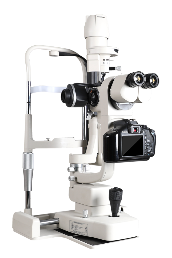
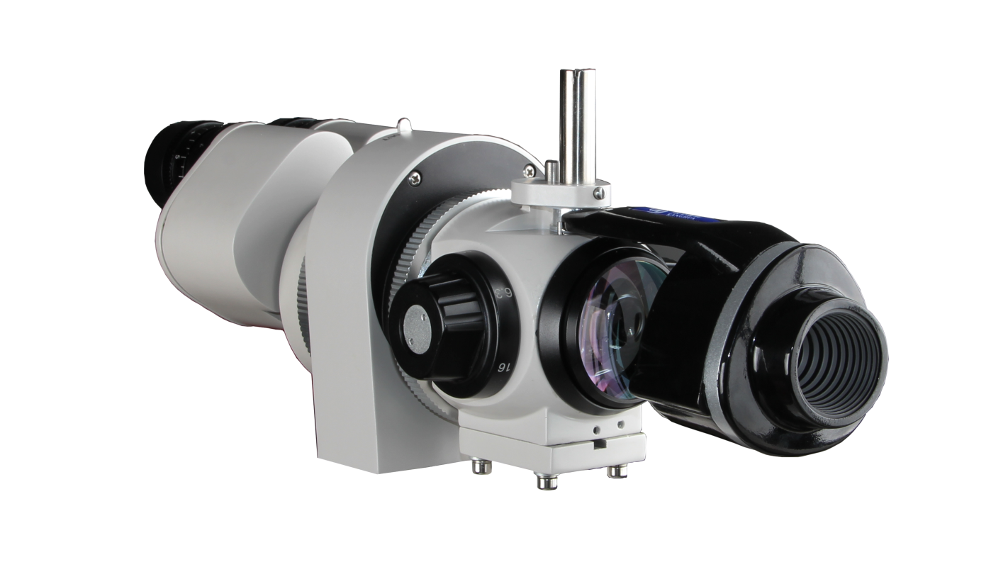
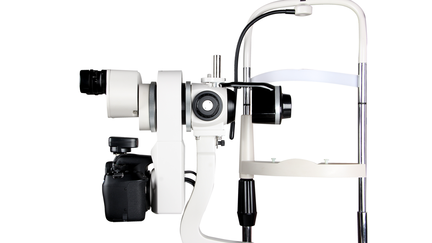
Optical microscopy combined with high-definition digital imaging to provide cell-level images. Non-irritating Infrared light illumination to ensure the accuracy of tear film measurement. One-stop inspection for anterior segment, operate easily and high-efficiency Optical resolution is 1900 lp/mm
Comprehensive Detection of Ocular Surface Microenvironment
The Dry Eye Analyzer integrates optical microscopy and high-definition digital imaging with non-invasive infrared illumination to observe and analyze the ocular surface in exceptional detail.
It enables real-time visualization and automatic measurement of key tear film parameters, including tear meniscus height (TMH), lipid layer thickness, and tear film break-up time (TBUT).
The system assists clinicians in accurately identifying tear film abnormalities and determining the type and severity of dry eye, ensuring precise and efficient diagnosis.
Comprehensive Diagnosis
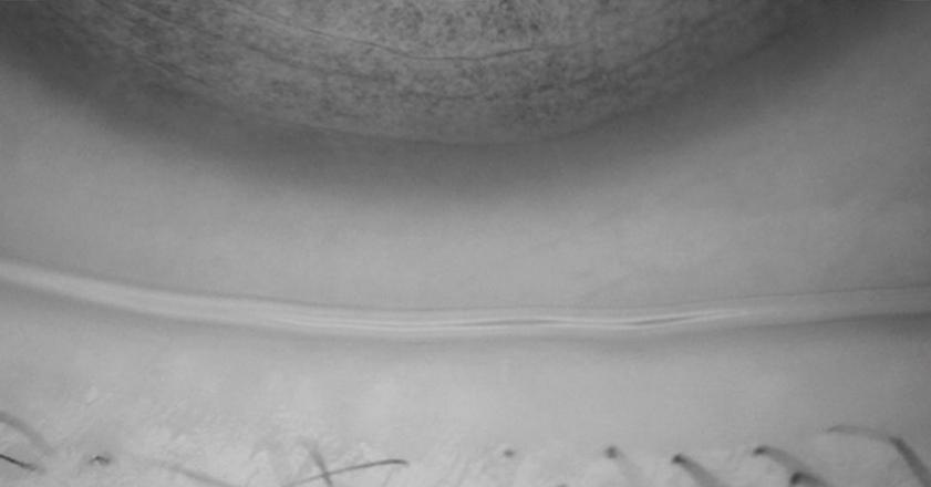
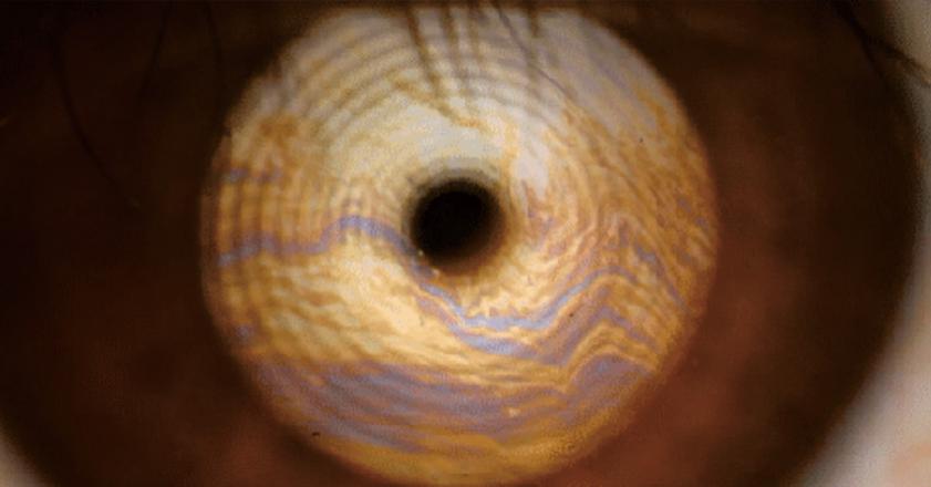
TEAR MENISCUS HEIGHT MEASUREMENT
Infrared light illumination is non-invasive and non-stimulating, and the tear secretion is no different from the normal state during measurement, ensuring the high data accuracy of TMH.
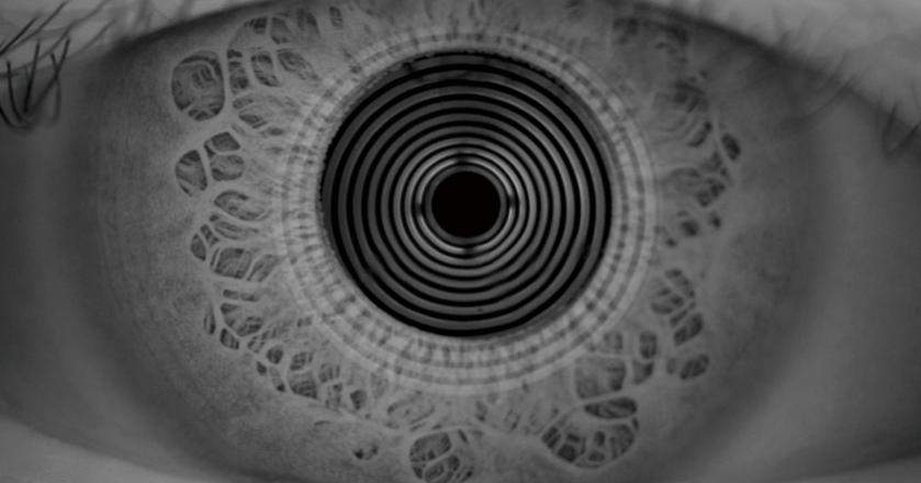
AUTO NIBUT
Infrared light illumination is different from the stimulation of visible light to the eyes of the subject. It not only ensures the successful completion of the test, but also does not interfere with the natural circulation of the tear film environment, and the test results are true and reliable.
LIPID LAYER EVALUATION
The unique structural design of the dry eye device maximizes the use of light effects, and presents the true color of the lipid layer on the basis of non-stimulating low-brightness lighting, thereby ensuring the accuracy of lipid layer thickness evaluation.
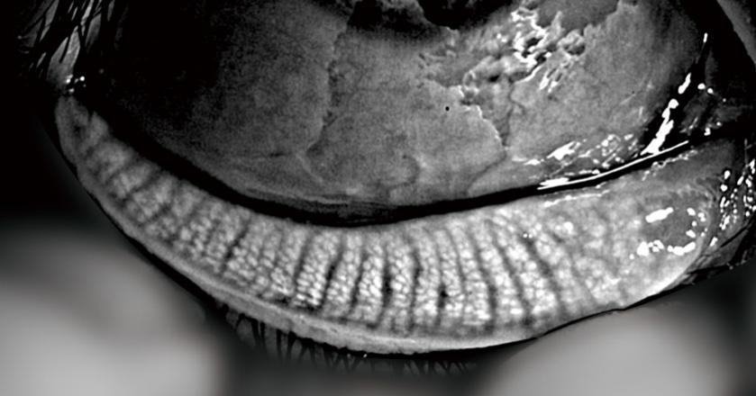
MEIBOMIAN GLAND DELETION
The acinar-level meibomian gland imaging, combined with infrared imaging and optical zoom, fully displays the details of the acinus, and supports clinicians in immediate treatment of the meibomian gland affected area. Comparison of standard templates to visually assess the degree of absence of meibomian glands.
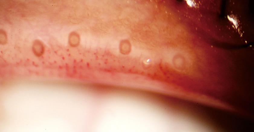
EYELID MARGIN ANALYSIS
The high-definition eyelid margin image can directly reflect the condition of the meibomian gland orifice, and it is clear at a glance whether it is blocked or not. Based on high-definition images and standard template comparison, the degree of changes in the meibomian gland orifice and the eyelid margin can be visually assessed.
REDNESS SCAN
The high-definition eyelid margin image can directly reflect the condition of the meibomian gland orifice, and it is clear at a glance whether it is blocked or not. Based on high-definition images and standard template comparison, the degree of changes in the meibomian gland orifice and the eyelid margin can be visually assessed.
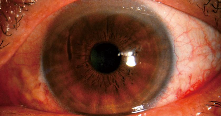
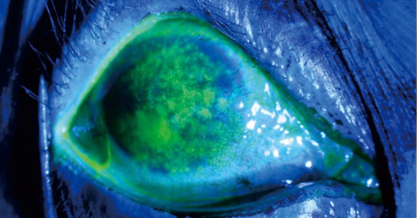
Corneal Staining
Reflects corneal epithelial integrity and assesses dry eye severity
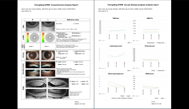
Comprehensive analysis report
Automatically generate reports according to the examination results without manually filling in. Illustrated report is easy to understand, operation process is simpler. Visible and intuitive report facilitate communication with patients.
FEATURES
ALL IN ONE
ALL IN ONE High efficiency equipment, Unique in the world, comprehensive inspection.
SALES LEAD
We have made the largest marke share in China, and also export to more than 100 countries and regions.
ACCURATE AND PROFESSIONAL
Non-invasive examination, accurate diagnosis of dry eye causes, assisting in formulating treatment plans.
INTELLIGENT
Fully automatic analysis of diagnostic results. Dry eye progress analysis report, showing dry eye treatment results for patients.
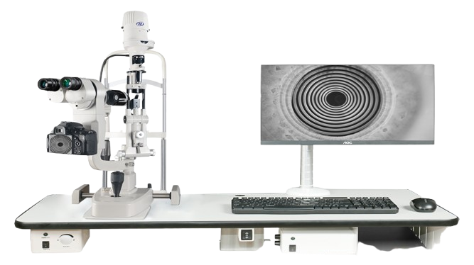
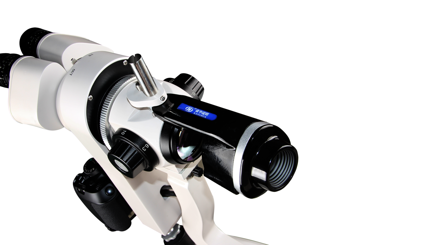
DRY EYE ANALYZER
Excellent optical microscopy, intelligent light conversion technology and high-definition digital imaging technology, through the hidden electronic control technology perfect union, carry on the classic ocular surface inspection platform. Cooperate with intelligent analysis software, not only can provide clinical dry eye examination with perfect high-definition images and accurate analysis results, can provide cells for the further study of ocular surface disease level super clear image and measurement data, can meet the needs of academic research.